Agent
There are two main causes of leg lesions in British finches.
Chaffinch papillomavirus is a virus in the Papillomaviridae family which can cause skin disease known as papillomatosis.
Cnemidocoptes spp. are mites which belong to the family Sarcoptidae that can cause skin disease known as cnemidocoptosis. In passerines (songbirds), C. jamaicensis and C. intermedius are the most common mite species; C. mutans typically affects poultry and C. pilae affects psittacine birds (such as pet budgerigars).
There are many colloquial names for these conditions, including “tassel foot” for papillomatosis and “mange” or “scaly foot” for cnemidocoptosis.
Species affected
In Great Britain (GB), both causes of leg lesions have been reported since the 1960s and the chaffinch (Fringilla coelebs) is by far the most frequently affected species. Brambling (Fringilla montifringilla), bullfinch (Pyrrhula pyrrhula), greenfinch (Chloris chloris) and goldfinch (Carduelis carduelis), as well as rook (Corvus frugilegus), Eurasian jackdaw (Corvus monedula), and carrion crows (Corvus corone), have also been reported with similar leg lesions, but only on rare occasions. More recently, a small number of dunnocks (Prunella modularis) have also been reported with such leg lesions, which were found to be caused by Cnemidocoptes spp. mites.
Pathology
Both papillomatosis and cnemidocoptosis can cause skin disease with similar appearance over the legs of finches.
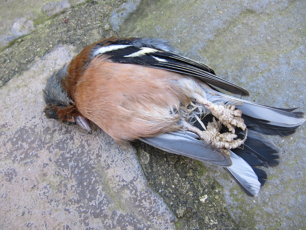
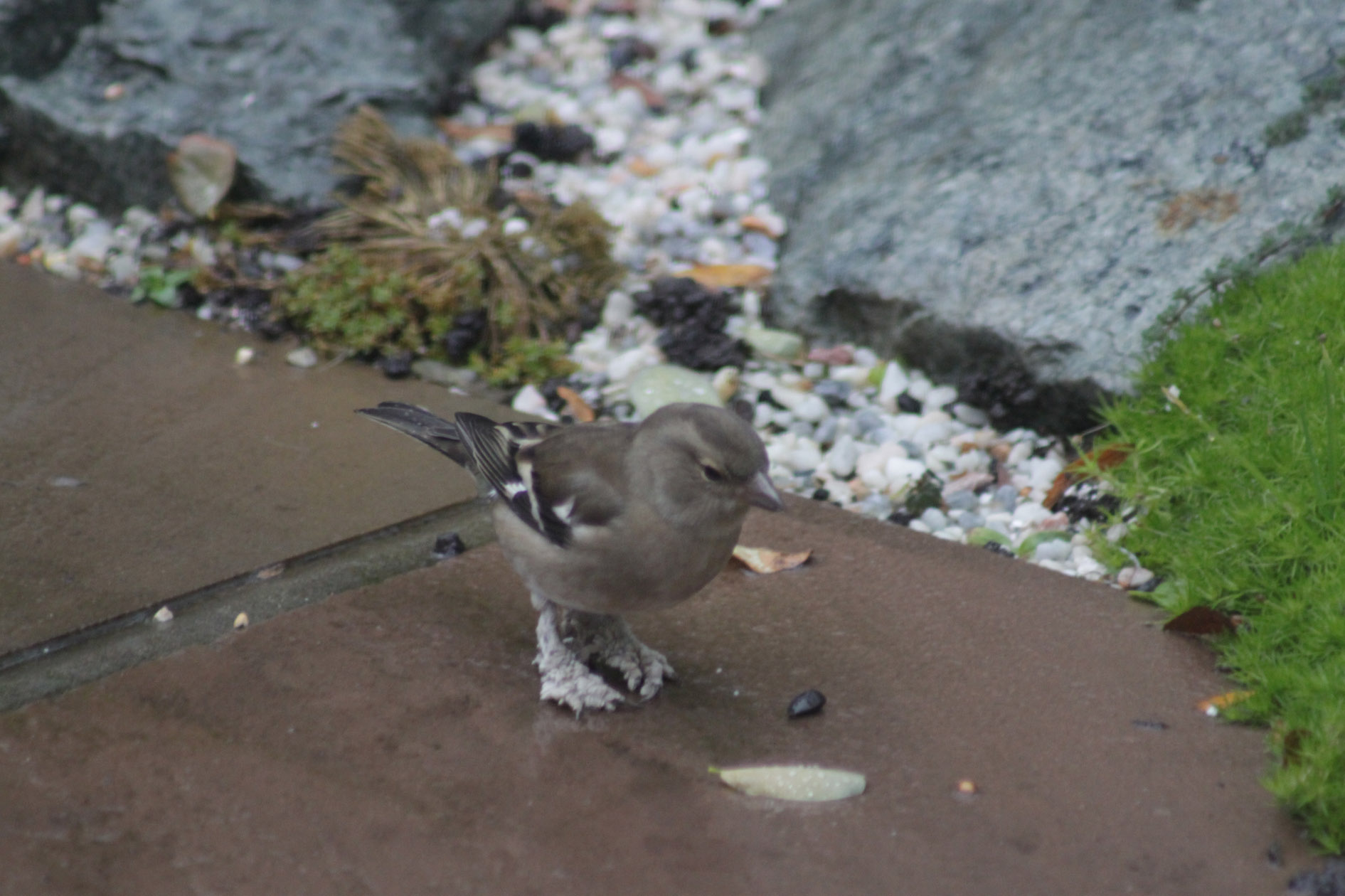
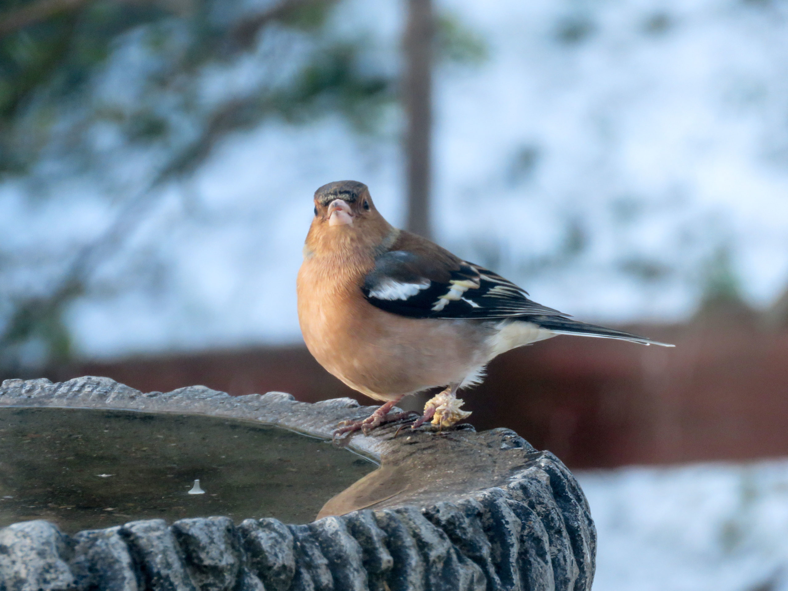
With papillomatosis, proliferative “spiky” or “tassel-like” lesions typically develop around the foot and digits (toes), but can spread higher up the leg.
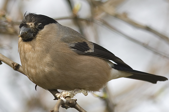
With cnemidocoptosis, excess white or grey-coloured skin growths can develop, involving the foot, digits or entire leg. On rare occasions, crusty, scab-like lesions can also occur on the face, in which case the condition is known colloquially as “scaly face”.
A recent study has found that mixed infection with both virus and mites is common and concluded that it is not possible to reliably discriminate between the two diseases based on the appearance of leg abnormalities alone.
Signs of disease
Chaffinches with leg lesions are most commonly bright and active, in normal body condition, and appear to be relatively unaffected by the condition. With both papillomatosis and cnemidocoptosis, skin abnormalities are thought to develop slowly over a period of weeks to months. Spontaneous recovery in a proportion of cases is believed to occur with papillomatosis, whilst the disease progression of cnemidocoptosis in wild birds is poorly understood.
Severe lesions can result in lameness or secondary bacterial infections, both of which can lead to debilitation, compromise welfare and lead to an increased susceptibility to predation and/or entanglement. One or both legs can be affected and digit loss can occur in some cases.
Figure 1. Chaffinch (Fringilla coelebs) with leg lesions. (Photo credit: Dave Sapp)
Figure 2. Bullfinch (Pyrrhula pyrrhula) with leg and facial skin lesions. (Photo credit: Dave Sapp)
Figure 3. Chaffinch (Fringilla coelebs) with leg lesions. (Photo credit: Brian Green)
Figure 4. Chaffinch (Fringilla coelebs) with leg lesions. (Photo credit: Alison Ritchie)
Disease transmission
Transmission of both papillomavirus and cnemidocoptic mites can occur via direct contact with affected birds or through indirect contact, for example at shared perches.
Disease patterns
Leg lesions in chaffinches have been reported in Great Britain and various other countries in continental Europe (including the Czech Republic, Germany, Sweden and the Netherlands) for multiple decades.
A recent study of leg lesions in chaffinches found that sightings of affected birds were reported from all regions across Great Britain. Each week, an average of 3 – 4% of BTO Garden BirdWatchers recording chaffinches saw a bird with leg lesions in their garden. These findings indicate that leg lesions are endemic (i.e. long-established) in the British chaffinch population. Whilst leg lesions were observed across the calendar year, there was a clear seasonal peak from November to March. The population of chaffinches in Great Britain is markedly increased during the winter months due to an influx of migratory birds from mainland Europe. The seasonal peak in occurrence of leg lesions is therefore likely to relate to the increased number of chaffinches in gardens, but may also be affected by climate, flocking and congregation at feeding stations, all of which could influence the likelihood of transmission.
Leg lesions usually only occur in a small proportion of chaffinches in a flock, with individual birds commonly affected. They are not a frequent cause of death but may compromise the welfare of affected birds: there is no evidence that they pose a conservation threat.
Risk to human and domestic animal health
Papillomaviruses are typically highly adapted to certain host species; therefore, chaffinch papillomavirus poses no known threat to human or other mammal species and is only likely to have the potential to cause disease in closely related wild or captive birds.
Cnemidocoptes mites are only known to infect birds; therefore, there is no known risk to human or other mammal health. However, garden birds in the UK may carry other infectious agents (for example Campylobacter, Chlamydia psittaci, Escherichia albertii and Salmonella bacteria) that can affect people and domestic animals.
We recommend following sensible hygiene precautions as a routine measure when feeding garden birds and handling bird feeders and tables. Following these rules will help avoid the risk of any infection transmitting to people and help safeguard the birds in your garden against disease.
-
Clean and disinfect feeders/ feeding sites regularly. Suitable disinfectants that can be used include a weak solution of domestic bleach (5% sodium hypochlorite) and other specially-designed commercial products (See Further information). Always rinse thoroughly and air-dry feeders before re-use.
-
Brushes and cleaning equipment for bird feeders, tables and baths should not be used for other purposes and should not be brought into the house, but be kept and used outside and away from food preparation areas.
-
Wear rubber gloves when cleaning feeders and thoroughly wash hands and forearms afterwards with soap and water, especially before eating or drinking. Avoid handling sick or dead birds directly. For instance, use disposable gloves or pick the bird up through an inverted plastic bag.
Risk to domestic animal health
The Cnemidocoptes species that typically infect songbirds (including finches) are different to the species of mite that commonly affect poultry: the risk of infection spreading from finches to poultry is therefore likely to be low. Whilst cage and aviary finches may be at risk of infection, transmission is unlikely since pet and wild birds would need to be in direct physical contact or share contaminated surfaces (e.g. perches) for this to occur.
Diagnosis
Whilst the appearance of finch leg lesions may be suggestive of either cause of the disease, it is not possible to reliably discriminate between the conditions based on visual inspection alone, and mixed infection with both agents may occur.
Since there is a significant ‘overlap’ in their appearance, diagnosis of papillomatosis and cnemidocoptosis infection in garden birds typically relies on post-mortem examination and additional laboratory tests (e.g. genetic tests, microscopic examination and electron microscopy).
If you wish to report finding dead garden birds, or signs of disease in garden birds, please visit www.gardenwildlifehealth.org. Alternatively, if you have further queries or have no internet access, please call the Garden Wildlife Health vets on 0207 449 6685.
Control
Whilst medicines are available for the treatment of cnemidocoptosis in captive birds, effective and targeted dosing of free-living birds is not possible. No medicine is available for the treatment of papillomatosis in wild or captive birds.
If several birds are affected in one area, general measures for control of disease in wild bird populations should be adopted:
-
Since infection with both agents is spread through direct physical contact between birds, or indirect contact (e.g. perches, feeding platforms), ensure optimal hygiene at garden bird feeding stations, including disinfection (as described above).
-
Feeding stations (such as bird tables and hanging feeders) encourage birds to congregate, sometimes in large densities, thereby increasing the potential for disease to spread between individuals when outbreaks occur. If many birds in your garden are affected, we recommend that you consider significantly reducing the amount you feed, or stop feeding for a period (2-4 weeks). The reason for this is to encourage birds to disperse, thereby minimising the chances of new birds becoming infected at the feeding station. Gradually reintroduce feeding, whilst continuing to monitor for further signs of ill health (See Further information).
Prevention
Following best practice for feeding garden birds is recommended to help control and prevent transmission of disease at feeding stations all year round (See Further information):
-
Routine good feeder hygiene. Clean away uneaten food and droppings before putting out fresh food and disinfect feeders/ feeding sites on a regular basis.
-
Provision of clean and fresh drinking water on a daily basis.
-
Provision of fresh food from reputable sources.
-
Rotate positions of feeders in the garden to avoid build-up of contamination in any one area and pay particular attention to clearing food remains that fall on the ground.
Further information
Best feeding practices should be followed at all times to help ensure that the birds visiting your garden remain healthy. More information can be found on the Garden Wildlife Health website www.gardenwildlifehealth.org.
Scientific publications
Seilern-Moy K, Heaver JP, Fernandez JR, Macgregor SK, John SK, Cunningham AA, Lawson B (2021) Atypical knemidokoptosis in two Dunnocks (Prunella modularis) in southern England. The Journal of Wildlife Diseases 57(2):467-70. doi.org/10.7589/JWD-D-20-00152
Lawson B, Robinson RA, Rodriguez-Ramos Fernandez J, John SK, Benitez L, Tolf C, Risely K, Toms MP, Cunningham AA, Williams RAJ (2018) Spatio-temporal dynamics and aetiology of proliferative leg skin lesions in wild British finches. Scientific Reports 8(1), p.1-12. doi.org/10.1038/s41598-018-32255-y
Pennycott TW (2016) Diseases of wild birds of the orders Passeriformes and Columbiformes – a review of conditions reported from the United Kingdom and an analysis of results from wild bird disease surveillance in Scotland 1994–2013. PhD Thesis, The University of Edinburgh, Edinburgh, UK, 417. https://era.ed.ac.uk/handle/1842/23001
Pence, D.B. Acariasis. (2008) In: Parasitic Diseases of Wild Birds. Atkinson C.T., Thomas, N.J., Hunter, D.B. (Eds..) Willey-Blackwell pp. 527-537. Erdélyi, K. (2012) Chaffinch Papilloma In: Infectious Diseases of Wild Mammals and Birds in Europe. Gavier-Widén D., Duff, J.P., Meredith, A. (Eds..) Wiley Blackwell p. 230-233.
Acknowledgements
Current funding for the GWH comes in part from Defra, the Welsh Government and the Animal and Plant Agency (APHA) Diseases of Wildlife Scheme (DoWS) http://apha.defra.gov.uk/vet-gateway/surveillance/seg/wildlife.htm; and from the Esmée Fairbairn Foundation, the Universities Federation for Animal Welfare and the Garfield Weston Foundation.
Disclaimer
This fact sheet was produced by Garden Wildlife Health (GWH) for information purposes only. The GWH will not be liable for any loss, damage, cost or expense incurred in or arising by reason of any person relying on information in this fact sheet.
Date of factsheet update:
